Loculated Pleural Effusion - Loculated Pleural Effusion Stock Image Image Of Hospital 132803315. Diffuse nodules and opacification in right lung with compressive atelectasis. Tube thoracostomy has variable success in the treatment of complex pleural effusions, with Nine of the 19 malignant effusions showed loculation (47%). Tell a friend about us, add a link to this page, or visit the webmaster's page for free fun content. Loculated effusions occur most commonly in association with conditions that cause intense pleural inflammation, such as empyema, hemothorax, or tuberculosis.
A pleural effusion is an unusual amount of fluid around the lung. Loculated effusions are difficult to confirm with chest radiograph, but ultrasound, computed tomography (ct), and even magnetic resonance imaging (mri) may be used to verify a localized collection of pleural fluid. This type of effusion is empyema unless proven otherwise. Activity intolerance related to acute pain secondary to pleural effusion, as evidenced by pain score of 10 out of 10, fatigue, disinterest in adls due to pain, dyspnea and orthopnea, verbalization of. Pleural effusion that is confined to one or more fixed pockets in the pleural space.
View Image from www.thoracicmedicine.org It is estimated that symptomatic pleural effusions affect more than 100,000 patients each year. Nine of the 19 malignant effusions showed loculation (47%). Sometimes in the setting of pleuritis, loculation of fluid may occur within the fissures or between the pleural layers (visceral and parietal). Normally, a small amount of fluid is present in the pleura. Tell a friend about us, add a link to this page, or visit the webmaster's page for free fun content. Surgical thoracostomy tube placement and radiologically guided catheter drainage are standard therapy for loculated pleural fluid collections. Tube thoracostomy has variable success in the treatment of complex pleural effusions, with Pleural effusion that is confined to one or more fixed pockets in the pleural space.
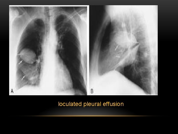
A loculated pleural effusion is the major radiographic hallmark of parapneumonic effusion or empyema (see fig.
Of the 22 transudates, eight showed a loculated pleural effusion (36%) compared with 45 of 78 exudates (58%). Icu patients cannot sit up and the effusion layers posteriorly. The pleura are thin membranes that line the lungs and the inside of the chest cavity and act to lubricate and facilitate breathing. Loculated effusions are difficult to confirm with chest radiograph, but ultrasound, computed tomography (ct), and even magnetic resonance imaging (mri) may be used to verify a localized collection of pleural fluid. The purpose of this study was to assess the value of intrapleural urokinase (uk) instillations in enhanc ing tube drainage of loculated, complex pleural effusions. 1 article features images from this case 20 public playlist includes this case Loculated effusions, defined as effusions that do not shift freely in the pleural space, occur when there are adhesions between the visceral and parietal pleura. Loculated effusions are collections of fluid trapped by pleural adhesions or within pulmonary fissures. Fluid levels in the right and left pleural cavities are often different, known as asymmetrical effusion. Pleural fluid is seen extending to the right oblique fissure. (vats) with lysis of adhesions is also a viable option for loculated effusions. Prior chest radiographs indicating that the blunting is a new finding also provide a good indicator of pleural effusion. Major imaging findings associated with complex loculated pleural effusions.
If you are struggling with chest pain that gets worse when you cough or inhale, chances. Pleural fluid is seen extending to the right oblique fissure. If it is clear that there are multiple loculations then it is wise to avoid delay and proceed directly to this procedure. Nursing care plan 3 nursing diagnosis: Fluid levels in the right and left pleural cavities are often different, known as asymmetrical effusion.
View Of Intra Pleural Tissue Plasminogen Activator And Deoxyribonuclease An Alternative Treatment Option For Pleural Infections In Specific Populations The Southwest Respiratory And Critical Care Chronicles from pulmonarychronicles.com Pleural effusion that is confined to one or more fixed pockets in the pleural space. Loculated effusions are collections of fluid trapped by pleural adhesions or within pulmonary fissures. Pleural effusions describe fluid between the two layer of tissue (pleura) that cover the lung and the lining of the chest wall. Causes of an exudative effusion are malignancy, infection, or inflammatory disorders such as rheumatoid arthritis. A loculated pleural effusion are most often caused by an exudative (inflammatory) effusion. Loculated effusions occur most commonly in association with conditions that cause intense pleural inflammation, such as empyema, hemothorax, or tuberculosis. Icu patients cannot sit up and the effusion layers posteriorly. Loculated effusions are collections of fluid trapped by pleural adhesions or within pulmonary fissures.
Empyema and large or loculated effusions need to be fo … at least 40% of all patients with pneumonia will have an associated pleural effusion, although a minority will require an intervention for a complicated parapneumonic effusion or empyema.
Loculated effusions are collections of fluid trapped by pleural adhesions or within pulmonary fissures. Pulmonology 16 years experience see below: Nine of the 19 malignant effusions showed loculation (47%). Complex, loculated pleural effusion visualized by us was defined as fibrin strands or septa floating inside the anechoic/hypoechoic pleural effusions along with presence of defined multiple pockets in the pleural cavity (10). Nursing care plan 3 nursing diagnosis: If it is clear that there are multiple loculations then it is wise to avoid delay and proceed directly to this procedure. Pleural effusions in the intensive care setting. The purpose of this study was to assess the value of intrapleural urokinase (uk) instillations in enhanc ing tube drainage of loculated, complex pleural effusions. Loculated effusions are difficult to confirm with chest radiograph, but ultrasound, computed tomography (ct), and even magnetic resonance imaging (mri) may be used to verify a localized collection of pleural fluid. It is estimated that symptomatic pleural effusions affect more than 100,000 patients each year. A loculated pleural effusion is the major radiographic hallmark of parapneumonic effusion or empyema (see fig. Empyema and large or loculated effusions need to be fo … at least 40% of all patients with pneumonia will have an associated pleural effusion, although a minority will require an intervention for a complicated parapneumonic effusion or empyema. Diffuse nodules and opacification in right lung with compressive atelectasis.
Pleural effusions describe fluid between the two layer of tissue (pleura) that cover the lung and the lining of the chest wall. The largest pocket of fluid is present posteriorly at the right lung base, with associated atelectasis and minor consolidation. Major imaging findings associated with complex loculated pleural effusions. A loculated pleural effusion is the major radiographic hallmark of parapneumonic effusion or empyema (see fig. Pleural fluid is seen extending to the right oblique fissure.
Pleural Disease Dr Nadya Ben Geweref Pleura Is from slidetodoc.com Malignant pleural effusion is a frequent complication of some common cancers. Pleural effusions in the intensive care setting. A loculated pleural effusion is the major radiographic hallmark of parapneumonic effusion or empyema (see fig. Pleural effusion, not elsewhere classified. Loculation most commonly occurs with exudative fluid, blood and pus. Of the 22 transudates, eight showed a loculated pleural effusion (36%) compared with 45 of 78 exudates (58%). Complex, loculated pleural effusion visualized by us was defined as fibrin strands or septa floating inside the anechoic/hypoechoic pleural effusions along with presence of defined multiple pockets in the pleural cavity (10). Icu patients cannot sit up and the effusion layers posteriorly.
Left pleural effusion with high density material at the posterior costophrenic angle.